Does exercise induce neuroplasticity?

Authors: Diana Sipos-Lascu, Oana-Maria Vanța
Keywords: aerobic exercises, neurotrophins, neuroimaging, brain plasticity
Does exercise induce neuroplasticity?
Is there a relationship between exercise and neuroplasticity? The term “neuroplasticity” refers to the nervous system’s ability to adapt its organizational structure to changing demands and circumstances. Neuroplasticity develops, for instance, when learning new abilities, after brain damage, and as a response to sensory deprivation. It is essential to promote neuroplasticity, as by modifying its structural and functional characteristics, the human brain adjusts to changing demands, which leads to learning and skill acquisition [1].
Convergent data from both human and animal studies showed that physical activity promotes neuroplasticity of some brain areas, increasing cognitive capabilities. According to animal research studies, the brain mechanisms by which physical exercise has a positive impact on cognition include an increase in:
- Neurogenesis
- Synaptogenesis
- Angiogenesis
- Release of neurotrophins [1].
Resistance training is a short-term, repetitive, monotonic, effortful, and voluntary muscular contraction that has been shown to improve gross motor function and maximal voluntary force. Neuroplastic changes in the central nervous system frequently follow improvements in motor ability [1].
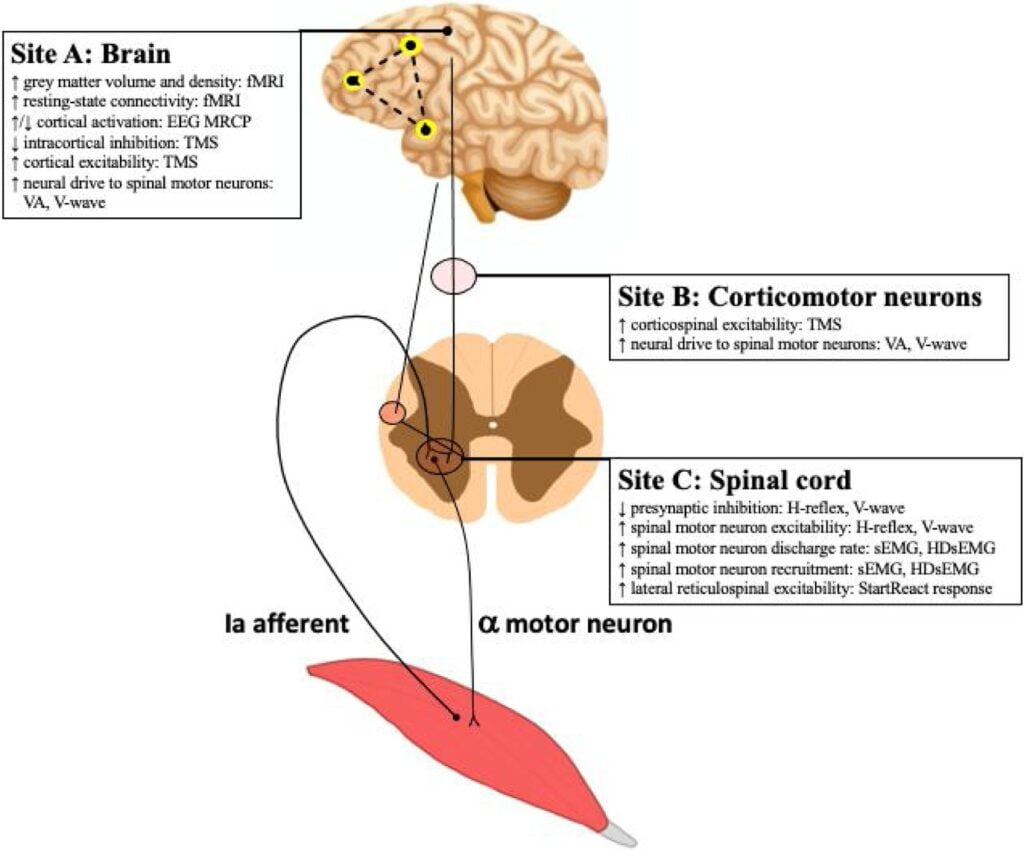
Some historical studies first proposed the concept of neuroplasticity, which describes structural and functional changes in particular brain and spinal circuits as a factor in the rapid initial rise in maximal voluntary muscle force in healthy humans following a period of resistance training (see Figure 1) [2].

Maximal voluntary muscle force is influenced by motor unit recruitment and the speed at which motor neurons release action potentials concerning measures of neuroplasticity in individual representative muscles of the agonist [2].
Which are the best exercises to promote neuroplasticity?
Aerobic exercise enhances cognitive and motor function by inducing brain changes that can be seen using molecular, cellular, and system-level neuroscience. It is a popular rehabilitation strategy for those who have suffered from neurological injury since it is a powerful way to induce neuroplasticity in the human brain.
Unfortunately, it is challenging to design the best exercise interventions for rehabilitation since it is not fully understood how aerobic exercise causes neuroplasticity. The Jenin El-Sayes review’s objective is to offer a thorough model describing the mechanisms of neuroplasticity brought on by persistent and intense exercise. Although the effects of chronic exercise on brain structure and function are well documented, it is still unclear how acute exercise causes neuroplasticity (see Table 1) [3].
| Long-Term Effects |
| MOLECULAR BDNF, VEGF, IGF-1 |
| CELLULAR Gliogenesis, Neurogenesis, Synaptogenesis, Angiogenesis |
| STRUCTURAL & FUNCTIONAL WMV, GMV, Receptor Activity, Neural Activity CBF |
| BEHAVIORAL Improved cognitive and motor function |
Table 1. The effects of continuous aerobic exercise on neuroplasticity. BDNF = brain-derived neurotrophic factor; IGF-1 = insulin-like growth factor 1; VEGF = vascular endothelial growth factor; WMV = white matter volume; GMV = gray matter volume; CBF = cerebellar blood flow.Available from [3].
Chronic aerobic activity elevates brain-derived neurotrophic factor (BDNF), insulin-like growth factor I (IGF-1), and vascular endothelial growth factor (VEGF) blood levels.
These elements encourage:
- angiogenesis
- synaptogenesis
- neurogenesis
- gliogenesis.
Increases in gray matter volume (GMV), white matter volume (WMV), and neuronal activity are most likely mediated by increases in neurogenesis. Increases in GMV and WMV are likewise most likely mediated by gliogenesis. Increases in brain activity and receptor activation observed after repeated aerobic exercise may be mediated by synaptogenesis. The rise in CBF may also be mediated by angiogenesis.
Improvements in cognitive and motor function are correlated with increases in:
- GMV
- WMV
- neuronal activity
- receptor activation.
How does chronic aerobic exercise induce neuroplasticity?
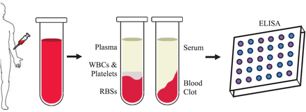
IGF-1, obtained from blood serum and plasma and measured using the ELISA test, is crucial for appropriate brain growth and maintenance (see Figure 2). Age appears to be a factor in the association between IGF-1 and regular exercise; cross-sectional studies demonstrate that young but not older adults exhibit a positive association between physical activity and IGF-1 levels. VEGF encourages the expansion of neural precursors and creates a vascular environment favorable for the development of neurons. Quantifying VEGF is similar to IGF-1 (see Figure 2). BDNF is responsible for increasing levels of IGF-1. The brain undergoes cellular changes from BDNF, IGF-1, and VEGF, including gliogenesis, neurogenesis, synaptogenesis, and angiogenesis [3].

What are angiogenesis, synaptogenesis, neurogenesis, and gliogenesis?
The process by which astrocytes, oligodendrocytes, and microglia are created is known as gliogenesis. Persistent exercise promotes gliogenesis, and rises in BDNF and IGF-1 may be responsible. The process through which new neurons are formed is known as neurogenesis. Chronic aerobic activity boosts neurogenesis, which could be brought on by an uptick in BDNF, IGF-1, and VEGF.
Sustained training increases synaptogenesis, which is the creation of connections between neurons. These adjustments are most likely caused by modifications in BDNF and IGF-1, BDNF controlling the development and proliferation of synapses. Continuous workout increases the process of angiogenesis, which represents the development of new blood vessels. Changes in cellular and molecular structure brought on by exercise are essential for causing changes in brain structure [3].
Which are the evidence of these adjustments?
Changes in cellular and molecular structure brought on by exercise are essential for causing changes in brain structure. Neuroimaging techniques differentiate the gray and white matter cortical regions altered by persistent exercise, and the increase in gray matter volume is at the level:
- hippocampus
- cerebellum
- basal ganglia
- cingulate cortices
- frontal cortices
- parietal
- occipital
- temporal
- insular cortices.
The white matter volume of the frontal, parietal, and occipital lobes likewise rises with chronic activity. Although most studies have concentrated on the elderly, the same effects are seen in both young and old adults. Improvements in neuronal integrity, a sign of healthy axons and myelin, and increased density and myelination induce changes in the gray and white matter. Increased gliogenesis, neurogenesis, and synaptogenesis—all of them being controlled by neurotrophic and growth factors—are most likely responsible for changes in brain structure. The correlation between rising BDNF levels and increased hippocampus volume lends credence to this.
Transcranial magnetic stimulation (TMS) (see Figure 3) is a noninvasive method that can be used to evaluate the short- and long-term changes in transcallosal communication and cortical neurotransmitter receptor activity that happen after acute and long-term exercise, respectively. Short-interval intracortical inhibition does not appear to be affected by physical activity levels. Exercise-induced increases in several brain areas are seen in functional magnetic resonance imaging activation in response to tasks that evaluate executive function. These include:
- the cingulate
- thalamus
- basal ganglia
- frontal
- temporal
- parietal
- occipital lobes
- insular
- motor cortices
- hippocampus.
In conclusion, chronic aerobic exercise changes behavior, which in this context is called altering cognitive and motor function. Following continuous exercise training, cognition improves due to molecular and cellular processes [3].

How does acute aerobic exercise induce neuroplasticity?
Acute aerobic exercise causes molecular, functional, and behavioral modifications like regular exercise does (see Table 2). However, unlike regular exercise, acute exercise is unlikely to result in cellular or structural changes. Acute exercise changes the amount of peripheral BDNF, IGF-1, and VEGF at the molecular level [3].
| Short-Term Effects |
| MOLECULAR VEGF, BDNF |
| FUNCTIONAL CBF, Glucose and O2 metabolism, NT concentrations, Neural Activity, Receptor Activity |
| BEHAVIORAL Improved cognitive and motor function |
Table 2. Acute aerobic exercise-induced neuroplasticity. BDNF = brain-derived neurotrophic factor; VEGF = vascular endothelial growth factor; NT = neurotransmitter; CBF = cerebellar blood flow; O2 = oxygen. Available from [3].
According to research, just one session of aerobic exercise boosts BDNF and VEGF. Acute exercise also raises the concentrations of neurotransmitters and metabolites. Additionally, the cerebellum, sensorimotor cortex, occipital cortex, and premotor areas experience increased glucose metabolism after engaging in acute aerobic activity. A single aerobic exercise session alone can change how the brain functions. Following acute exercise, receptor activity is changed as measured by TMS. Via resting-state functional MRI, acute exercise improves connectivity between brain areas. Moreover, acute exercise can alter behavior and enhance cognitive and motor performance [3]. Some factors can impact neuroplasticity induced by aerobic sessions like:
- biological sex
- genetic variations
- high levels of estradiol
- the individual’s basal fitness level [3].
What is the conclusion?
- There are important mechanisms behind neuroplasticity brought on by aerobic exercise.
- Aerobic workout increases white matter volume by upregulating gliogenic and neurogenic processes, whereas gliogenesis, neurogenesis, and synaptogenesis enhance gray matter volume.
- Exercise-induced improvements in cerebrovascular function most likely bring on structural adjustments.
- Increased brain activation and communication efficiency result from anatomical changes by exercise.
- How effective aerobic activity is for neuroplasticity depends on some individual factors.
- Future research should thoroughly define how these factors regulate neuroplasticity and develop new and better techniques by understanding how these parameters affect the induction of neuroplasticity by aerobic exercises.
For more information on neurorehabilitation, visit:
- The importance of physical activity in neurorecovery
- Intrinsic Mechanisms of Acupuncture in Ischemic Stroke RehabilitationRobotic neurorehabilitation for the upper limb – new insights
- Efficacy of placebo in managing pain for neurological disorders
We kindly invite you to browse our Interview category: https://efnr.org/category/interviews/. You will find informative discussions with renowned specialists in the field of neurorehabilitation.
References
- Höttin K, Röder B. Beneficial effects of physical exercise on neuroplasticity and cognition. Neuroscience and Biobehavioral Reviews. 2013. DOI: 10.1016/j.neubiorev.2013.04.005
- Hortob’agyi T, Granacher U, Fernandez-del-Olmo M, Howatson G, et al. Functional relevance of resistance training-induced neuroplasticity in health and disease Neuroscience and Biobehavioral Reviews. 2021. DOI: 10.1016/j.neubiorev.2020.12.019
- El-Sayes J, Harasym D, Turco CV, Locke MB, Nelson AJ. Exercise-Induced Neuroplasticity: A Mechanistic Model and Prospects for Promoting Plasticity. The Neuroscientist 2019, Vol. 25(1) 65–85. DOI: 10.1177/1073858418771538


